Wilting Disease Diagnosis: Distinguishing Between Fungal Wilt and Environmental Stress
Examine vascular changes closely-fungal wilts show distinct browning xylem with pathogen-specific patterns like Fusarium’s reddish streaks or Verticillium’s black dots. Wilting progresses gradually in fungal cases, starting at leaf bases and spreading upward, while environmental stress causes rapid, uniform drooping. Check for tylose formations and compare symptom timelines to differentiate between these issues. As you explore further, you’ll learn how to combine vascular analysis with leaf patterns and testing for accurate diagnoses.
Notable Insights
- Examine vascular discoloration patterns – fungal wils show specific streaks and tyloses, while environmental stress causes uniform wilting without internal changes.
- Assess symptom progression timeline – fungal infections develop gradually over weeks, whereas environmental stress causes rapid wilting within days.
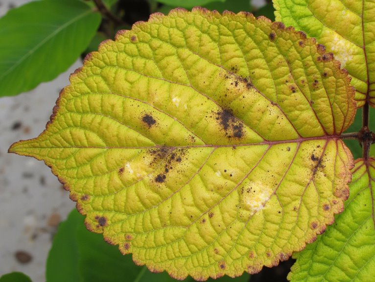
- Analyze leaf curling and necrosis – asymmetrical curling from the base up suggests fungal blockage, while uniform browning indicates water deficiency.
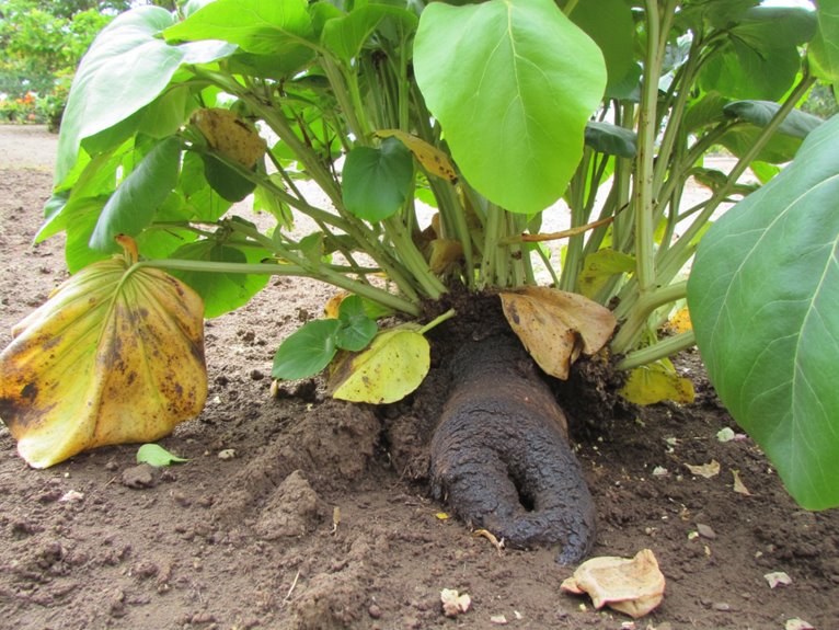
- Check for root/vascular discoloration – Phytophthora shows stem rot, while drought stress lacks internal changes entirely.
- Confirm with laboratory tests – microscopic examination reveals pathogen-specific structures like spores or microsclerotia in vascular tissues.
Identifying Vascular Discoloration in Fungal Wilt
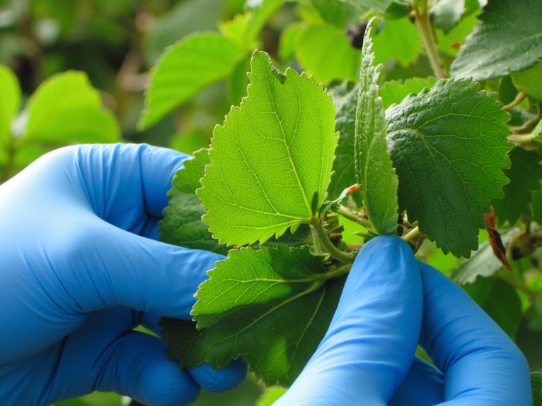
When examining plants showing wilt symptoms, looking for vascular discoloration can be both revealing and rewarding for growers, as this subtle but significant indicator often points directly to fungal pathogens beneath the surface.
Mastering vascular observation techniques helps identify fungal colonization indicators like browning xylem vessels or tylose formations blocking water flow. These changes reflect pathogen activity within plant tissues rather than just environmental stress.
Vascular inspection reveals fungal colonization through browning vessels and tylose blockages, offering early disease detection beyond surface symptoms.
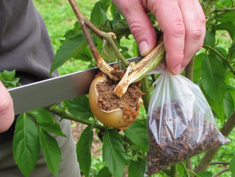
Fusarium infections show reddish-brown streaks and salmon-colored spores, while Verticillium produces black microsclerotia in vascular tissue. Using sharp tools to cut stems reveals these patterns clearly.
Growers should check for uniform discoloration around the vascular ring or localized streaks indicating pathogen spread. Noting these signs early lets you isolate affected plants and apply targeted treatments before wilting becomes permanent.
Practical applications include checking corms of cyclamen for Fusarium infections or observing v-shaped leaf lesions with Verticillium. Maintaining proper NPK ratios through fertilization can help strengthen plant resistance to these vascular diseases.
Combining visual inspection with lab testing confirms diagnoses accurately. Developing these skills improves your ability to protect crops from fungal wilt diseases effectively.
Progression of Wilting Symptoms: Fungal Vs Environmental
As you watch a plant wither, distinguishing between fungal and environmental wilting reveals essential insights into what’s happening beneath the surface.
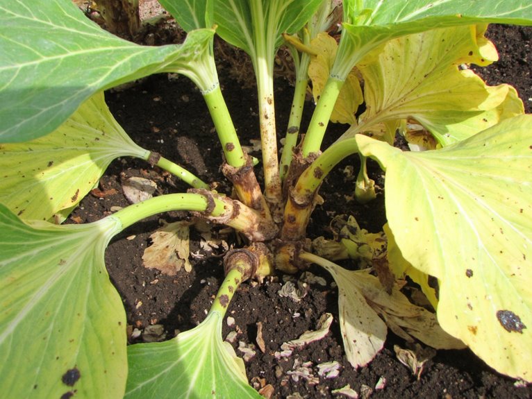
Fungal wilts like Verticillium typically begin with gradual symptoms mid-season or during heat/drought stress, starting on older leaves and progressing section by section. Environmental stress causes more uniform wilting that may reverse with improved conditions.
The symptom timeline helps: fungal infections typically worsen over weeks to seasons, while environmental issues can cause rapid collapse within days.
Stress impact varies too – fungal pathogens exacerbate wilting from heat or drought, but environmental fixes alone might restore health if the root cause is addressed.
Leaf Necrosis and Specific Curling Patterns
Let’s take a closer look at how leaves turn brown and curl – key clues that can help diagnose whether the problem stems from a fungus or environmental stress.
Fungal wilts like Fusarium create distinct leaf curling patterns, such as epinasty, where young leaves droop downward due to blocked water flow. Vascular blockage is a critical factor in these patterns, often resulting in asymmetrical curling that starts at the base of the leaf and progresses upward.
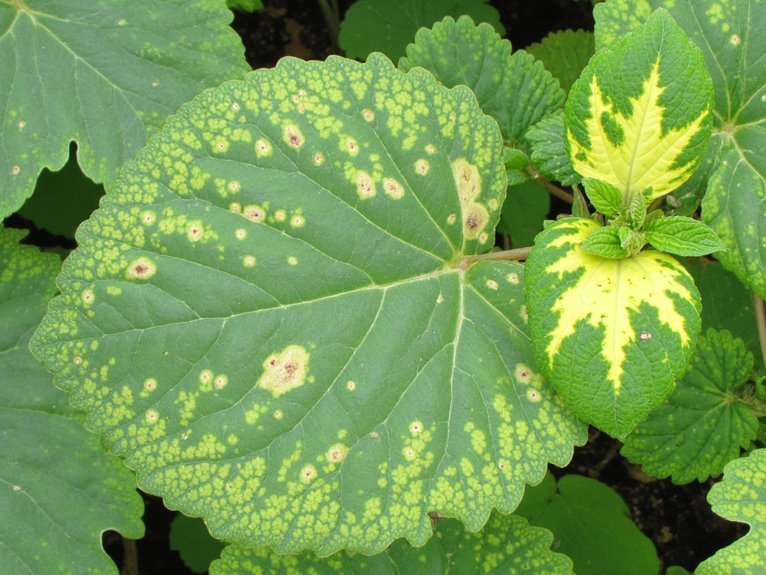
Necrosis assessment reveals critical differences: fungal infections often start at edges and spread inward, creating crispy, dry dead areas. Environmental stress causes more uniform browning.
When you examine these symptoms together, the pattern tells a story – asymmetrical necrosis with specific curl’s suggests vascular issues, while generalized rolling points to abiotic factors.
Diagnostic Testing for Fusarium and Verticillium Wilts
You’ll want to look at vascular discoloration first-it’s like the plant’s own red flag saying something’s wrong down inside its tubes.
Then you’ll need lab tests, whether that’s looking at fungal bits under a microscope or doing those fancy DNA checks that can pin down exactly which wilt pathogen you’re dealing with.
The key is knowing when to combine symptoms with science, not just go by looks alone.
Vascular Discoloration Analysis
Even though vascular discoloration might seem like a simple sign to look for, it’s actually quite telling when it comes to diagnosing fungal wilts in plants. Examining vascular discoloration patterns and conducting thorough wilted plant inspection reveals critical clues about the pathogen at play.
| Disease | Discoloration Color | Inspection Focus |
|---|---|---|
| Fusarium | Brown | Stem streaks |
| Verticillium | Medium to dark brown | Sapwood blotches |
| Oak Wilt | Brown | Bark-peeled vascular rings |
Look for temperature and pH correlations, as these factors influence discoloration intensity. Sampling thumb-thick branches with symptomatic leaves yields the best results for analysis.
Laboratory Confirmation Methods
While laboratory confirmation might seem challenging, it’s an essential step in accurately diagnosing fungal wilts in plants.
PCR techniques amplify DNA from pathogens like Fusarium and Verticillium, even when present in small amounts. Microscopic identification examines spore structures on specialized media to distinguish species.
Spectroscopy applications, like Raman spectroscopy, detect molecular changes before visible symptoms appear. Pathogenicity assays confirm isolates actually cause disease by inoculating plants.
Diagnostic validations guarantee these methods work reliably across different environments and hosts. Together, these approaches provide a thorough understanding of the pathogen, guiding targeted management strategies for growers.
Symptom Differentiation Techniques
After confirming a pathogen through laboratory techniques like PCR and microscopy, growers need to distinguish between similar fungal diseases that cause wilting.
Understanding symptom patterns helps guide intervention strategies and improve symptom management. Fusarium shows yellow vascular streaks in stems, while Verticillium creates dark brown or black discoloration.
Check stem cuts diagonally for distinct vascular signs. Verticillium often causes unilateral symptoms like red leaf discoloration or partial wilting that may resolve temporarily – a key clue for treatment timing.
Monitor progress using wilt severity scales (0-3) to track changes. Environmental stress can worsen both diseases, so evaluating growing conditions is essential for targeted solutions.
Recognizing Bacterial Wilt Signs and Symptoms
As you monitor your plants for stress, keeping an eye on subtle changes can make all the difference, especially with bacterial wilts that often start quietly.
Early detection of bacterial wilts in plants hinges on noticing quiet shifts-like subtle leaf drooping and color changes-that signal underlying infection.
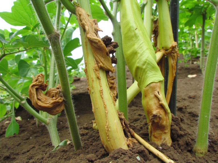
Wilting appears five days after infection, with partial leaves drooping during heat and full leaves yellowing, necrotizing between veins. Vascular symptoms show as stems develop light tan discoloration progressing to dark streaks and hollow interiors.
Cut stems reveal bacterial ooze-sticky slime or milky beads-that protects pathogens and confirms active infection. Unlike fungal causes, bacterial wilts produce this distinct exudate and show rapid decline with brown petioles.
Inspect vascular patterns and test for ooze to distinguish these symptoms from environmental stress.
Differentiating Phytophthora Root Rot From Wilting Diseases
Though wilting can hint at many problems, Phytophthora root rot stands out because it often starts below ground-only revealing its presence when plants show signs above.
Above-ground symptoms like yellowing and sparse foliage are common to many issues, but Phytophthora symptoms reveal themselves through root decay and stem discoloration.
Fibrous roots rot first, turning black and brittle, while larger roots develop distinct brown lesions. This differs from drought stress, which doesn’t cause internal discoloration.
Management focuses on drainage improvement and soil sterilization to combat this persistent pathogen.
Frequently Asked Questions
How to Differentiate Pathogen-Caused Wilting From Physical Damage?
You need to examine symptom comparison closely – pathogen-caused wilting shows gradual, asymmetric patterns with vascular streaks when stems are split, while physical damage causes abrupt, uniform wilting without discoloration.
Root analysis reveals the truth: fungal wilt shows rot or lesions, whereas mechanical trauma displays broken, crushed, or desiccated roots with no biological signs.
Look for pathogen evidence like mycelium or spores too.
With these distinctions clear, you’ll confidently diagnose and nurture your plants back to health.
Role of Soil Conditions in Fungal Wilt Progression?
Soil moisture levels directly impact pathogen survival and root health-both overly wet and droughty soils can favor infection.
Maintain consistent moisture without waterlogging, as compacted or acidic soils worsen symptoms.
Build resilient root systems with balanced nutrients and proper structure to outsmart pathogens naturally.
Resistant Plant Varieties for Fusarium/Verticillium?
You’re looking for plant varieties that stand a better chance against Fusarium and Verticillium wilts-good news!
Tomato hybrids like Aligote F1 and Celebrity Plus F1, plus strawberries like Portola and UC Monarch, show strong disease resistance through smart breeding strategies.
These resistant plants don’t just fight one pathogen but often battle multiple races and related diseases too.
Choosing these varieties helps you manage wilt risks naturally, reducing your reliance on chemicals while boosting yields.
Start by checking which resistant cultivars work best for your growing conditions!
Wilting Confused With Nutrient Deficiencies?
You’re right to wonder about wilting confused with nutrient deficiencies – they often look similar!
Nutrient imbalance can cause yellowing, stunted growth, and even necrotic spots that mimic disease symptoms.
But there’s good news: microbial interactions in the soil can help differentiate these issues. For instance, fungal pathogens often cause wilting with distinct root discoloration, while nutrient deficiencies show specific patterns like interveinal chlorosis.
Testing soil and understanding your garden’s microbiome will give you the best chance to diagnose accurately.
Incubation Period for Fungal Wilt Diseases?
It varies widely depending on the plant, pathogen strain, and environment. For watermelon, symptoms can show up in as little as two weeks or as late as thirteen weeks.
Disease symptoms often start subtly-yellowing leaves, stunted growth-before progressing to defoliation.
Knowing this timeframe helps you spot issues earlier and take action before plants suffer irreversible damage.
On a final note
Distinguishing fungal wilt from environmental stress starts with examining vascular discoloration – that telltale yellowing in stems only professionals might notice. While both cause wilting, fungal diseases often progress suddenly and affect entire branches, whereas stress-related symptoms typically improve with care changes. Don’t despair if your plants look sick – with careful inspection and possibly lab testing, you can pinpoint the cause and save your garden.